An exercise stress test evaluates how the heart responds when it works harder, revealing blockages or electrical problems that remain hidden at rest. During the test, patients walk on a treadmill while specialized equipment continuously tracks heart rate, rhythm, blood pressure, and electrical activity (ECG). This test is performed under constant medical supervision, making it extremely safe, with serious complications occurring in less than 1 in 10,000 cases. This effective diagnostic method assists in diagnosing coronary artery disease, evaluating the effectiveness of applied treatment, and determining safe activity levels, providing critical information about the patient’s heart health.
Overview
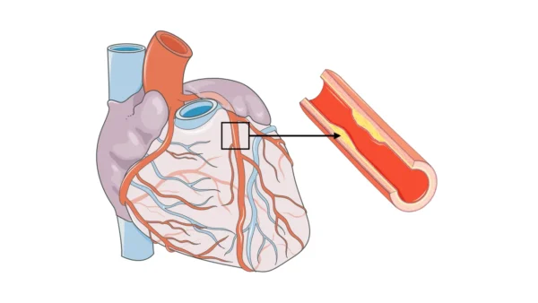
The exercise cardiac stress test, often called a treadmill test or exercise electrocardiogram (ECG), assesses how your heart and circulatory system function during increased physical activity. When your heart needs more oxygen, healthy arteries widen to meet the demand. However, if your coronary arteries are narrowed by plaque buildup, they fail to deliver enough oxygen during exertion. This vital mismatch between oxygen supply and demand creates detectable changes on the monitoring equipment.
The basic test involves the patient walking on a motorized treadmill or pedaling a stationary bike while connected to an ECG machine that continuously records the heart’s electrical activity. Blood pressure is measured frequently. The exercise intensity is gradually increased, typically every two to three minutes, following standardized protocols such as the Bruce protocol. This progression continues until the patient reaches their calculated target heart rate, develops concerning symptoms, or shows signs that require the test to stop.
The entire procedure, including preparation and recovery, usually takes 45–60 minutes, although the active exercise phase is typically only 8–15 minutes. The test is highly safe; serious complications, such as a heart attack or dangerous rhythm problem, occur in less than 1 in 10,000 tests. Experienced medical personnel continuously monitor the patient and are prepared to manage any issues immediately.
Why You Might Need One
An exercise stress test is recommended in several situations to gather vital information about heart health:
- Diagnosing Coronary Artery Disease (CAD) is the most common reason. If a patient experiences chest pressure, tightness, or pain—particularly during activity—the test helps determine if blocked arteries are responsible.
- Shortness of breath that seems out of proportion to the activity level helps distinguish between cardiac and non-cardiac causes.
- Assessing known heart disease following a heart attack, stenting, or bypass surgery shows whether new blockages have developed or if existing treatments remain effective.
- Guiding treatment and activity in patients with heart failure or certain rhythm problems evaluates exercise capacity, assists in determining prognosis, and establishes safe limits for daily physical activity or return to sports.
- Screening high-risk individuals who have multiple risk factors (diabetes, high blood pressure, high cholesterol, or a strong family history) and are planning to start a vigorous exercise program helps screen for hidden problems.
Important Note: Patients experiencing severe chest pain at rest or showing signs of an active heart attack are not admitted for a stress test; they are immediately directed to the emergency room for urgent evaluation and treatment.
Preparing for the Test
Proper preparation is crucial for accurate results and patient safety during the stress test:
- Clothing the patient should wear comfortable clothes and athletic shoes suitable for brisk walking or running. Women should wear a bra, as electrodes are attached to the chest.
- Medication instructions the physician provides specific guidance on which medications to take or hold; certain medications like beta-blockers or calcium channel blockers may need to be held for 24–48 hours before the test, as these drugs lower heart rate and can mask signs of disease; no medication should ever be stopped without the explicit instruction of the physician.
- Eating and drinking a light meal should be consumed 2 to 4 hours before the test, avoiding heavy meals; most importantly, all caffeine (coffee, tea, soda, chocolate) must be avoided, and the patient must not smoke for at least 3 to 4 hours prior to the test, as these substances affect heart rate and test interpretation.
- Inhalers patients who use an inhaler for asthma or lung disease should bring it with them.
- Physical limitations the patient must immediately inform the staff if they have joint problems, balance issues, or anything that prevents adequate walking on the treadmill, as alternative pharmacological stress tests may be more appropriate.
What Happens During the Test
The procedure begins with preparation. A technician cleans and places 10 or more sticky electrode patches on the patient’s chest, connecting them to the ECG machine. A blood pressure cuff is placed on the arm. Resting heart rate, blood pressure, and ECG are recorded to establish a baseline.
Throughout the exercise, the medical staff continuously monitors several factors:
- ECG changes the staff observes for ST segment depression, a specific pattern that strongly suggests the heart muscle is not receiving adequate blood flow (iskemi) during stress.
- Blood pressure response the systolic pressure is expected to rise progressively; a failure of pressure to rise adequately, or a drop in pressure during exercise, suggests severe cardiac problems.
- Symptoms the patient is frequently asked about chest discomfort, shortness of breath, dizziness, or other symptoms.
- Rhythm problems dangerous heart rhythm changes, such as ventricular tachycardia, are monitored.
The recovery phase begins immediately when the patient stops exercising. The patient walks slowly for a few minutes to cool down. ECG and blood pressure monitoring continue for at least 5–10 minutes during recovery, as some concerning changes only appear during this period.
Interpreting the Data
The stress test provides multiple pieces of crucial information used to assess heart health:
- Exercise capacity the duration of exercise and the level reached indicate overall cardiovascular fitness; stopping very early or failing to adequately raise the heart rate suggests significant underlying heart issues or severe deconditioning.
- Ischemia evidence (ECG) the most critical finding is the ST segment depression pattern, which is strong evidence that narrowed coronary arteries cannot deliver enough oxygen to the heart muscle when stressed.
- Blood pressure dynamics the rate at which blood pressure rises and falls indicates the health of the vascular system; an abnormal or dropping blood pressure response suggests severe cardiac problems.
- Heart rate recovery how quickly the heart rate drops in the first minute after exercise is a powerful predictor of long-term outcomes; a decrease of less than 12 beats per minute suggests a higher cardiovascular risk.
- Symptom reproduction if the test reproduces the patient’s typical chest discomfort accompanied by relevant ECG changes, it strongly confirms the diagnosis of angina from coronary artery disease.
Advanced Stress Tests
Basic stress tests provide valuable information, but enhanced versions offer more definitive, visual assessment:
- Stress echocardiography adds ultrasound images of the heart taken immediately before and after exercise; the staff looks to see if areas of the heart supplied by potentially narrowed arteries contract weakly or not at all when stressed, providing visual confirmation of inadequate blood flow.
- Nuclear stress tests involve injecting a small amount of a radioactive tracer; special cameras capture images showing blood flow to the heart muscle, comparing rest and exercise images; this method is highly accurate for detecting areas of poor blood flow.
- Pharmacologic stress tests utilize medications for patients unable to exercise due to physical limitations (such as arthritis); these drugs simulate the effects of exercise, thereby allowing the imaging portion of the test to be performed.
Results and What They Mean
Understanding the test results is key to determining the patient’s next steps:
- Normal (negative) test signifies the patient exercised adequately, reached the target heart rate, had no symptoms, and showed no concerning ECG or imaging abnormalities; this result is highly reassuring, suggesting that the coronary arteries are open and supplying adequate blood flow at the time of testing.
- Abnormal (positive) test indicates significant ECG changes, especially if accompanied by symptoms, suggesting the presence of coronary artery disease; abnormal imaging further confirms this diagnosis.
- Inconclusive test occurs if the patient was unable to exercise adequately to reach the target heart rate, limiting the test’s ability to fully stress the heart.
Following an abnormal stress test, additional testing often follows. Coronary angiography—an invasive test where catheters and dye visualize the arteries directly—is typically the next step to definitively locate the blockage and determine whether treatment requires medications, stenting, or bypass surgery.
Limitations and Accuracy
While stress tests are valuable, they are not perfect.
- False positives occur when the test suggests coronary disease but arteries are actually normal, sometimes happening in patients taking certain medications or in women.
- False negatives occur when the test appears normal despite significant coronary disease being present; this happens when narrowings are not severe enough to limit blood flow during the exercise level achieved.
- The test only evaluates blood flow at one point in time meaning a normal result does not eliminate the possibility of disease developing later.
Enhanced imaging reduces both false positive and false negative rates compared to ECG monitoring alone. Despite these limitations, stress tests remain a valuable screening tool.
Key Points
To assist in quickly recalling the most critical information and steps, here are the key points about the exercise stress test:
- The exercise stress test evaluates heart performance under physical activity, aiming to reveal problems that are not apparent at rest.
- The primary purpose of the test is to diagnose coronary artery disease in symptomatic individuals, evaluate exercise capacity in known heart patients, and confirm the effectiveness of current treatment regimens.
- Safety the procedure is highly safe, with serious events occurring in less than 1 in 10,000 cases, due to continuous medical monitoring.
- Crucial preparation includes wearing athletic shoes, eating lightly 2–4 hours prior, and following specific instructions to hold certain heart medications for up to 48 hours before the test (under physician guidance).
- The test provides vital data on exercise capacity, blood pressure response, heart rhythm changes, and, most importantly, ECG changes (ST segment depression) suggesting inadequate blood flow (ischemia).
- Enhanced tests (Stress Echo or Nuclear Stress Test) provide visual evidence of poor blood flow and are more accurate than the basic ECG test alone.
- A positive (abnormal) result often leads to further definitive testing, such as coronary angiography, to determine the exact location and severity of any blockages.
- Collaboration with the cardiologist is essential for the patient to effectively use the test results, as they form a crucial part of the overall strategy for managing heart health.
Reference: Stress Testing