Your heart is a muscular pump about the size of your fist that beats roughly 100,000 times every day, pumping blood throughout your entire body. It sits in the center of your chest, slightly tilted to the left, behind your breastbone. Every heartbeat pushes oxygen-rich blood to every organ, tissue, and cell in your body, delivering the oxygen and nutrients they need to function. At the same time, your heart collects blood that has delivered its oxygen and pumps it to your lungs to pick up fresh oxygen.
Overview
Your heart has four chambers working together like a coordinated pumping system. Think of it as two pumps side by side, each with two chambers.
The right side receives blood returning from your body after delivering oxygen to tissues. This blood is low in oxygen and needs to go to your lungs. The right side pumps this blood to your lungs where it picks up fresh oxygen and releases carbon dioxide you exhale.
The left side receives oxygen-rich blood returning from your lungs. This blood is bright red and ready to deliver oxygen throughout your body. The left side pumps this blood to every part of your body—your brain, muscles, organs, skin, everywhere.
Each side has an upper chamber called an atrium that receives blood, and a lower chamber called a ventricle that pumps blood out. The upper chambers are smaller and thinner because they just collect blood and pass it down. The lower chambers are larger and more muscular because they do the hard work of pumping blood out to your lungs and body.
Four valves act like one-way doors, ensuring blood flows in the correct direction and doesn’t leak backward. These valves open to let blood through, then snap shut to prevent backflow.
Your heart beats in a coordinated rhythm. Both upper chambers fill with blood, then squeeze together, pushing blood into the lower chambers. Then both lower chambers squeeze together powerfully, pushing blood out to your lungs and body. The whole cycle takes less than a second. Then it repeats, over and over, roughly 60-100 times per minute when you’re resting.
When you exercise or get stressed, your heart beats faster and more forcefully, delivering more oxygen to meet your body’s increased needs. When you sleep, it slows down since your body needs less.
The Four Chambers
Your heart’s four chambers each have specific jobs.
- The right atrium is the upper chamber on the right side. It receives blood returning from your entire body through large veins. This blood has already delivered its oxygen to tissues and is dark bluish-red. The right atrium collects this blood and passes it down to the right ventricle.
- The right ventricle is the lower chamber on the right side. It receives blood from the right atrium above it. When it contracts, it pumps blood through your pulmonary arteries to your lungs. The right ventricle doesn’t need to pump very hard because your lungs are close by.
- The left atrium is the upper chamber on the left side. It receives oxygen-rich blood returning from your lungs through pulmonary veins. This blood is bright red because it’s full of fresh oxygen. The left atrium collects this blood and passes it down to the left ventricle.
- The left ventricle is the lower chamber on the left side. It’s the most muscular chamber because it does the hardest work—pumping blood to your entire body. When it contracts, blood shoots out through your aorta, the body’s largest artery, and travels to every part of your body from head to toe. The left ventricle’s thick muscular walls generate enough force to push blood through thousands of miles of blood vessels.
The walls separating the right and left sides are solid—blood doesn’t normally cross between sides inside your heart. The right side handles low-oxygen blood going to the lungs. The left side handles oxygen-rich blood going to the body. They work simultaneously but handle different blood.
The Four Valves
Valves ensure blood flows in one direction through your heart, preventing backflow.
- The tricuspid valve sits between the right atrium and right ventricle. When the right atrium fills with blood, this valve opens and blood flows down into the right ventricle. When the right ventricle squeezes, the valve snaps shut, preventing blood from flowing back up. The name comes from having three flaps.
- The pulmonary valve sits at the exit of the right ventricle where blood leaves to go to your lungs. When the right ventricle contracts, this valve opens and blood flows into the pulmonary arteries heading to your lungs. After the ventricle finishes contracting, the valve closes, preventing blood from flowing backward into the heart.
- The mitral valve sits between the left atrium and left ventricle. When the left atrium fills with oxygen-rich blood from your lungs, this valve opens and blood flows down into the left ventricle. When the left ventricle contracts, the valve closes tightly, preventing backflow. This valve has two flaps.
- The aortic valve sits at the exit of the left ventricle where blood leaves to go to your body. When the left ventricle contracts powerfully, this valve opens and blood shoots out into your aorta. After contraction, the valve closes, preventing blood from flowing back into the heart.
All four valves open and close in perfect coordination with each heartbeat. When working properly, they open fully to allow easy blood flow and close completely to prevent leaks. Problems occur when valves become too narrow to open fully or too loose to close completely.
How Blood Flows
Understanding blood flow through your heart helps you see how everything works together.
- Blood returning from your body enters the right atrium through large veins. This blood has delivered oxygen throughout your body and now needs fresh oxygen.
- From the right atrium, blood flows down through the tricuspid valve into the right ventricle.
- The right ventricle contracts, closing the tricuspid valve and opening the pulmonary valve. Blood flows through the pulmonary valve into pulmonary arteries going to your lungs.
- In your lungs, blood picks up oxygen and releases carbon dioxide. This happens in tiny air sacs where blood vessels and air come very close together.
- Oxygen-rich blood returns from your lungs through pulmonary veins to the left atrium.
- From the left atrium, blood flows down through the mitral valve into the left ventricle.
- The left ventricle contracts powerfully, closing the mitral valve and opening the aortic valve. Blood shoots out through the aortic valve into your aorta, the body’s largest artery.
- From your aorta, blood travels through progressively smaller arteries reaching every part of your body. Blood delivers oxygen to tissues, then collects in veins that eventually return to the right atrium, completing the cycle.
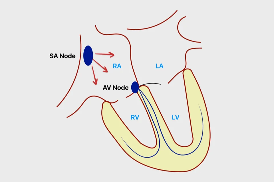
The Heart’s Electrical System
Your heart beats because of electrical signals that coordinate its contractions. This system works automatically without you thinking about it.
- The natural pacemaker sits in the right atrium. This small area of specialized cells generates electrical signals automatically, typically 60-100 times per minute when you’re resting. This is your heart’s built-in pacemaker.
- Each electrical signal spreads across both atria, causing them to contract together. Blood squeezes from the atria down into the ventricles.
- The signal then passes through a relay station that briefly delays it. This delay allows the atria to finish contracting before the ventricles start.
- After this delay, the signal travels down specialized pathways to both ventricles, causing them to contract together powerfully. Blood pumps out to your lungs and body.
- Then the cycle repeats. The pacemaker fires another signal and the whole process happens again.
- Your heart rate adjusts automatically based on your needs. When you exercise, nervous system signals and hormones tell your pacemaker to speed up. When you relax or sleep, it slows down. This happens automatically.
- Problems can occur anywhere in this electrical system. The pacemaker might fire too slowly, too quickly, or irregularly. Extra electrical pathways might exist that shouldn’t. Areas might become irritable and fire signals when they shouldn’t. These problems cause arrhythmias.
Coronary Arteries
Your heart muscle needs its own blood supply to function. Coronary arteries provide this.
- Three main coronary arteries wrap around the outside of your heart, branching into smaller vessels that penetrate deep into the heart muscle. These arteries deliver oxygen-rich blood to every part of your heart muscle.
- Your heart muscle works constantly, so it needs continuous oxygen delivery. Coronary arteries stay open and blood flows through them between heartbeats.
- When coronary arteries become narrowed by plaque buildup, blood flow becomes restricted. Your heart muscle doesn’t get enough oxygen, particularly during exertion when it needs more. This causes chest pain called angina.
- If a coronary artery becomes completely blocked—usually when plaque ruptures and a blood clot forms—the heart muscle supplied by that artery doesn’t receive any oxygen. Muscle tissue begins dying within minutes. This is a heart attack.
- Keeping coronary arteries healthy by controlling cholesterol, blood pressure, diabetes, maintaining healthy weight, not smoking, and staying active prevents these problems.
The Cardiac Cycle
Each heartbeat follows a pattern called the cardiac cycle.
- Diastole is the relaxation phase. Your heart muscle relaxes and chambers expand. Blood flows from the atria down into the ventricles. The ventricles fill with blood. This phase takes slightly longer than systole.
- Systole is the contraction phase. First the atria squeeze, pushing any remaining blood down into the ventricles. Then the ventricles contract powerfully. Valves between atria and ventricles snap shut. Valves at the ventricle exits open. Blood shoots out to your lungs and body.
- After systole, the heart relaxes again, beginning another diastole. The cycle repeats continuously.
- When doctors measure blood pressure, they’re measuring the pressure in your arteries during systole and diastole. The top number is systolic pressure—the pressure when your heart contracts. The bottom number is diastolic pressure—the pressure when your heart relaxes.
What You Hear
The sounds your heart makes—the “lub-dub” heard through a stethoscope—come from valves closing.
- The “lub” is the first heart sound. It happens when the tricuspid and mitral valves close as the ventricles begin contracting. This is usually louder and longer than the second sound.
- The “dub” is the second heart sound. It happens when the aortic and pulmonary valves close after the ventricles finish contracting. This is usually shorter and higher-pitched.
- Abnormal sounds called murmurs indicate turbulent blood flow, often from valve problems. Leaky valves create swishing sounds as blood flows backward. Narrowed valves create harsh sounds as blood is forced through tight openings.
- Doctors can tell a lot about your heart’s health just by listening carefully to these sounds.
Key Points
- Your heart is a muscular pump with four chambers—two upper chambers called atria that receive blood, and two lower chambers called ventricles that pump blood out.
- The right side receives blood from your body and pumps it to your lungs to get oxygen. The left side receives oxygen-rich blood from your lungs and pumps it to your entire body.
- Four valves act like one-way doors, opening to let blood through and closing to prevent backflow. Problems occur when valves don’t open fully or don’t close completely.
- Blood flows in a specific path: body to right atrium, to right ventricle, to lungs, to left atrium, to left ventricle, then out to the body, completing the cycle.
- Your heart beats because of electrical signals generated by a natural pacemaker. These signals spread in an organized pattern, causing coordinated contractions.
- Your heart rate adjusts automatically—faster when you exercise or are stressed, slower when you rest or sleep.
- Coronary arteries wrapped around your heart supply oxygen to the heart muscle itself. Blockages in these arteries cause heart attacks.
- Each heartbeat has two phases—diastole when the heart relaxes and fills with blood, and systole when it contracts and pumps blood out.
- Your heart beats roughly 100,000 times daily, pumping about 2,000 gallons of blood through your body.
Reference: Heart work