Overview
An ECG is a simple test that records the electrical signals your heart makes each time it beats. When your heart squeezes to pump blood, an electrical wave moves through it. The ECG picks up these waves and shows them as lines on a screen or printed paper.
To do this, small sticky patches called electrodes are placed on your chest, arms, and legs. These electrodes sense tiny electrical changes on your skin and send them to the ECG machine, which turns them into a heart tracing.
A normal ECG has a familiar pattern with three main waves: the P wave, the QRS wave, and the T wave. The P wave shows the upper chambers of your heart squeezing. The QRS wave shows the lower chambers squeezing. The T wave shows the heart resetting before the next beat. By looking at these waves, doctors can tell if your heart is beating too fast, too slow, or irregularly.
There are different types of ECGs. A resting ECG is done while you lie still. A stress ECG records your heart while you walk on a treadmill or use a bike. A Holter monitor is a small, portable ECG you wear for 1–2 days to record your heart during normal activities. An event monitor records your heart only during symptoms or when it detects something unusual.
The test is completely safe. It does not send electricity into your body— it only listens to what your heart is already doing.
Why You Might Need One
Doctors order ECGs for many reasons. If you have chest pain, an ECG can show whether your heart is under stress or having a heart attack. During a heart attack, part of the heart does not get enough blood, and the ECG pattern changes in a way doctors can recognize.
If you feel your heart racing, fluttering, or skipping beats—called palpitations—an ECG can show if you have an irregular rhythm such as atrial fibrillation or other rhythm problems. Since some irregular rhythms come and go, you may need a longer test like a Holter monitor.
Symptoms like dizziness, fainting, trouble breathing, or extreme tiredness can also be caused by heart rhythm problems. An ECG can help find out why.
Before surgery, an ECG gives doctors a quick look at how your heart is working so they can plan anesthesia safely. Some medicines can affect heart rhythm, so an ECG may be done before starting these medications.
People with risk factors for heart disease—such as high blood pressure, diabetes, high cholesterol, smoking, or family history—may have ECGs as part of routine check-ups.
Athletes in certain sports may get ECGs to screen for rare heart conditions that could be dangerous during intense exercise.
Before the Test
There is almost nothing you need to prepare for an ECG. You can eat and drink normally. Keep taking your usual medications unless your doctor says otherwise.
Wear loose clothing so the technician can place the electrodes easily. Avoid lotions, oils, or powders on your chest because they make it harder for the electrodes to stick.
Sometimes a small area of chest hair may need to be shaved to help the electrodes stay in place.
If you are doing a stress ECG, wear comfortable clothes and shoes for exercise. Don’t eat a large meal right before the test. Bring your inhaler if you use one.
Tell the technician if you have allergies to sticky adhesives.
The Test
For a resting ECG, you will lie down on a table. The technician cleans small spots on your skin and attaches about ten electrodes—six on your chest and four on your arms and legs. These electrodes connect to the ECG machine with thin wires.
You will be asked to lie still and breathe normally. The recording takes less than a minute, though the whole process takes about ten minutes. You won’t feel anything—no shocks, no pain.
The ECG shows twelve different views of your heart’s activity, each from a slightly different angle. This helps the doctor see if any part of your heart is not working correctly.
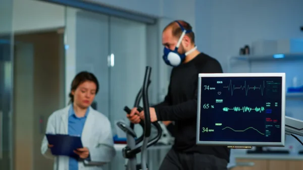
For a stress ECG, the electrodes stay on while you walk or jog on a treadmill or pedal a bike. Your heart rate, rhythm, and blood pressure are watched during exercise and recovery.
A Holter monitor or event monitor records your heart for a day or more. You wear the device under your clothes and go about your normal activities.
What to Expect During the Test
In a resting ECG, the only sensations you’ll notice are the cool alcohol wipes and the slight stickiness of the electrodes. The room might be a bit cold, but you can usually have a blanket.
If you’re having a stress ECG, you’ll feel your heart and breathing speed up as you exercise. This is normal. Tell the team right away if you feel chest pain, dizziness, or anything unusual.
Wearing a Holter monitor can feel a bit awkward at first, but most people get used to it quickly.
After the Test
For a resting ECG, the electrodes are gently removed—this may feel like peeling off a bandage. You can return to normal activities right away.
After a stress ECG, you’ll rest for a short time while your heart rate returns to normal. You may feel tired afterward, especially if you are not used to exercising.
When a Holter monitor test is done, you return the device and the recordings are analyzed by your doctor.
Understanding Your Results
A doctor interprets the ECG by examining the shapes, timing, and patterns of the waves. A normal ECG shows a steady heartbeat and normal electrical activity.
Abnormal results can show many different problems, such as irregular rhythms, signs of a past or current heart attack, thickened heart muscle, or problems caused by low or high levels of certain minerals in the blood.
Medication effects can also appear on the ECG. Your doctor will explain any findings and whether more tests or treatment are needed.
Living with ECG Monitoring
If you need to wear a monitor for a longer period, you can usually continue most of your everyday activities. Keep the device dry and record any symptoms you have in a diary.
Avoid bumping or pulling on the wires. Sleep as usual; many people find wearing a T-shirt helps keep the wires in place.
When to Call Your Doctor
After a routine ECG, you don’t usually need to call your doctor unless you have new symptoms. Seek medical help right away if you have chest pain, severe shortness of breath, fainting, or rapid heartbeats.
If you are wearing a monitor and you have severe symptoms, get emergency help—but keep the device on if possible so the rhythm can be recorded.
Call your doctor if the electrode sites become very irritated or if you haven’t received your results within the expected time.
If your ECG shows something abnormal, follow all recommended tests or treatments. An ECG is just a snapshot, so ongoing symptoms may require more testing such as longer monitoring or imaging.
You may also like to read these:
Arrhythmias: Symptoms, Causes, and Treatment
Reference: Electrocardiogram