Your heart beats because of electrical signals traveling through it in precise patterns. When these signals go wrong, your heart can beat too fast, too slow, or irregularly, causing palpitations, dizziness, fainting, or even sudden cardiac arrest. An electrophysiology study—or EP study—is a detailed test that maps your heart’s electrical system from the inside to find exactly what’s causing rhythm problems. A doctor threads thin, flexible catheters through blood vessels to your heart and uses them to record electrical activity from multiple locations simultaneously, creating a detailed picture of how signals travel.
Overview
An electrophysiology study examines your heart’s electrical system by recording electrical activity from inside your heart chambers. This provides far more information than a standard electrocardiogram, which only records from the surface of your body.
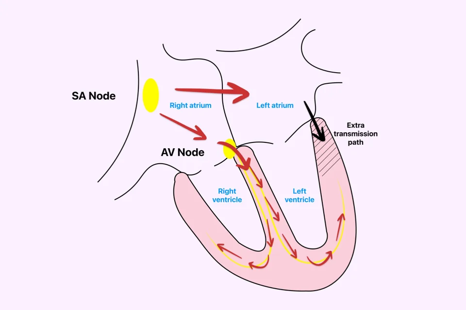
Your heart’s electrical system normally starts in the upper right chamber and spreads in an organized pattern, making your heart beat steadily at 60-100 times per minute. Problems occur when extra pathways form between chambers, certain areas become irritable and fire signals too rapidly, or normal pathways conduct abnormally.
Surface electrocardiograms sometimes show these problems but often can’t pinpoint exactly where they originate. EP studies provide this precision by placing recording electrodes directly on heart tissue.
During the study, catheters with multiple electrodes are positioned in different heart chambers, recording electrical activity from many locations simultaneously. Doctors can see exactly when each area activates, how quickly signals travel, and whether abnormal signals or pathways exist.
The study also involves pacing—delivering small electrical impulses through the catheters to make your heart beat at different rates or to trigger abnormal rhythms. This helps doctors understand what’s causing your symptoms.
EP studies diagnose conditions including supraventricular tachycardia, atrial fibrillation, atrial flutter, ventricular tachycardia, and unexplained fainting. Many studies combine diagnosis with treatment—if an abnormality is found, ablation is often performed immediately to fix the problem.
Why You Might Need This
Several situations lead doctors to recommend EP studies.
- Palpitations that haven’t been diagnosed despite wearing monitors warrant investigation. If monitors haven’t captured your episodes of rapid heartbeat, an EP study can trigger them in a controlled setting.
- Unexplained fainting, particularly in young people, requires evaluation. EP studies identify whether your heart’s electrical system is prone to life-threatening rhythms.
- Survivors of sudden cardiac arrest need EP studies to understand why their hearts stopped and determine whether an implantable defibrillator is needed.
- Known rhythm problems that need treatment benefit from EP studies. For supraventricular tachycardia, the study pinpoints where the problem originates for targeted ablation. For atrial fibrillation, it maps your heart before ablation.
- Very slow heart rates with unclear causes require investigation to determine whether you need a permanent pacemaker.
- People with weak heart muscle or certain genetic conditions face higher risks for dangerous rhythms. EP studies assess this risk and guide decisions about implantable defibrillators.
Preparing for the Study
Proper preparation ensures the study provides accurate information safely.
- Your doctor reviews all medications. Some rhythm medications must be stopped several days before the study so doctors can see your heart’s natural behavior. You receive specific instructions—never stop medications without guidance.
- Don’t eat or drink anything after midnight before your study.
- Blood tests check kidney function, blood counts, and electrolyte levels.
- If you take blood thinners, your doctor provides specific instructions about whether to continue or stop them.
- Arrange for someone to drive you home and stay with you. You’ll receive sedation and can’t drive.
- Plan for a possible hospital stay—most people go home the same day, but if ablation is performed you might stay overnight.
What Happens During the Study
Knowing the step-by-step process helps you understand what to expect, even though you’ll be sedated during much of it.
- You’re brought to the electrophysiology lab and given sedation through an IV—enough to make you drowsy but able to respond to instructions. Some studies use general anesthesia where you’re completely asleep.
- Small punctures are made in veins in your groin, sometimes in your neck. Local anesthetic numbs these areas completely. Thin tubes called sheaths are inserted through these punctures.
- Multiple catheters are threaded through the sheaths to your heart using X-ray guidance. Each catheter has electrodes that record electrical activity. You don’t feel catheters moving through vessels.
- Once positioned, doctors record your heart’s electrical activity from multiple locations. They measure how quickly signals travel and whether abnormal pathways exist.
- Small electrical impulses are then delivered through the catheters, pacing your heart at different rates. This isn’t painful—you might notice your heart beating faster but it doesn’t hurt. The pacing triggers your abnormal rhythm so doctors can study exactly where it starts and how it travels.
- If an abnormality is found that can be fixed with ablation, it’s often performed immediately. The ablation catheter destroys the small area causing problems using radiofrequency energy or extreme cold.
- After ablation, doctors repeat testing to confirm the abnormality is gone. They try to trigger abnormal rhythms again—if they can’t, the ablation succeeded.
- The study typically takes 1-3 hours, longer if ablation is performed.
- Catheters are removed and pressure is applied to puncture sites for 10-30 minutes to stop bleeding.
After the Study
Recovery begins immediately with careful monitoring.
- You’re moved to recovery. If groin access was used, you must lie flat for 4-6 hours to prevent bleeding. This is uncomfortable but essential.
- Nurses check your vital signs and puncture sites frequently. As sedation wears off, you become more alert. You can eat and drink once you’re not nauseated.
- Your doctor discusses findings—what rhythm problems were found, whether ablation was performed, and what happens next.
- Most people having diagnostic-only studies go home the same day. If ablation was performed, you might stay overnight.
- If ablation fixed your rhythm problem, medications for that rhythm might be stopped. You receive clear instructions about medication changes.
Recovery at Home
Recovery after EP studies is relatively quick, though some precautions are necessary.
- The first 24 hours involve rest. Avoid strenuous activity and heavy lifting.
- Groin puncture sites will bruise—this is normal and fades over 1-2 weeks. Keep areas clean and dry. Shower after 24 hours.
- Watch for problems: large or growing swelling, persistent bleeding, increasing pain, or leg numbness all require immediate attention.
- If you had ablation, you might feel tired for several days. Some chest discomfort or extra heartbeats for a few days usually resolve on their own.
- Most people return to work within 2-3 days, longer if ablation was performed.
- Don’t drive for 24 hours due to sedation effects.
Understanding Your Results
EP study findings guide diagnosis and treatment decisions.
- Normal findings mean your heart’s electrical system functions properly without abnormal pathways or irritable areas.
- Supraventricular tachycardia findings reveal extra pathways or re-entry circuits, usually successfully treated with ablation. Success rates exceed 95%.
- Atrial fibrillation studies show where abnormal signals from pulmonary veins trigger episodes, guiding ablation strategy.
- Atrial flutter studies map the circuit causing rapid atrial rhythm. Ablation typically cures this immediately.
- Ventricular tachycardia findings identify dangerous fast rhythms in lower chambers. Some can be fixed with ablation; others require implantable defibrillators.
- Conduction system problems show abnormally slow signal transmission, helping decide if you need a pacemaker.
Potential Complications
EP studies are generally safe, but complications can occur.
- Bleeding at puncture sites is most common. Minor bruising affects nearly everyone. Larger collections of blood occasionally occur but usually resolve without treatment.
- Blood vessel damage can happen but is uncommon.
- Heart perforation is rare but serious—catheters can puncture heart walls, requiring immediate treatment.
- Abnormal rhythms triggered during the study usually stop on their own or with medications.
- Stroke can occur if blood clots travel to the brain, though protective measures minimize this risk.
- Damage to normal electrical pathways during ablation can occur, potentially requiring a permanent pacemaker.
- Death from EP studies is extremely rare, occurring in less than 1 in 1,000 procedures.
Key Points
- Electrophysiology studies examine your heart’s electrical system by recording activity from inside your heart chambers, providing far more information than surface electrocardiograms.
- Multiple catheters are threaded through veins to your heart to record electrical activity, map signal travel, and trigger rhythm problems in a controlled setting.
- You’re sedated during the 1-3 hour procedure. Most people don’t remember much about it.
- EP studies diagnose supraventricular tachycardia, atrial fibrillation, atrial flutter, ventricular tachycardia, and causes of unexplained fainting or very slow heart rates.
- Many studies combine diagnosis with treatment—if an abnormality is found, ablation can often fix it immediately.
- Preparation includes stopping certain medications as directed, fasting after midnight, and arranging transportation.
- Recovery is quick. Most people go home the same day and return to normal activities within 2-3 days.
- Complications are uncommon but include bleeding, blood vessel damage, or heart perforation. Serious complications occur in less than 1% of procedures.
- Successfully ablated rhythm problems often never recur, curing many people permanently.
- Work with your electrophysiologist to understand your results and develop an appropriate treatment plan.
You may also like to read these:
Reference: Electrophysiological Study