An echocardiogram is an ultrasound of your heart—the same technology used to see babies during pregnancy, but focused on your heart. A technician places a probe on your chest that sends sound waves through your skin. These waves bounce off your heart’s structures and return to create moving images on a screen. The test shows your heart’s chambers, valves, and walls in real-time as they beat, revealing how well your heart pumps, whether valves are leaking or narrowed, if chambers are enlarged, and whether any structural abnormalities exist. It’s completely painless and takes about 30-45 minutes.
Overview
An echocardiogram uses high-frequency sound waves to create moving pictures of your heart. The technology is similar to ultrasounds used during pregnancy—completely safe and painless.
A small handheld device called a transducer is placed on your chest. It sends sound waves through your skin that bounce off heart structures and return to the device. A computer converts these returning sound waves into detailed moving images displayed on a screen.
The test shows your heart’s four chambers, four valves, major blood vessels, and the muscular walls. Doctors can see everything moving in real-time—chambers filling and emptying, valves opening and closing, walls thickening and relaxing with each heartbeat.
Several measurements are made during the test. The size of each chamber is measured to see if any are enlarged. Wall thickness is measured to detect thickening or thinning. How much blood the left ventricle pumps with each beat—called ejection fraction—is calculated. Valve function is assessed to see if they’re leaking or narrowed. Blood flow patterns are visualized to detect abnormal flows.
Echocardiograms diagnose many conditions. Heart failure is confirmed by seeing weak pumping or stiff walls. Valve problems are identified by watching how valves open and close and measuring blood flow through them. Heart attacks are detected by seeing wall segments that don’t move properly. Congenital heart defects show up as abnormal structures or connections. Fluid around the heart is easily visible.
Types of Echocardiograms
Several variations exist depending on what information doctors need.
- Transthoracic echocardiogram is the standard type. The transducer is placed on your chest wall. This provides good images for most purposes and is completely non-invasive.
- Transesophageal echocardiogram provides more detailed images by placing the transducer on a flexible tube passed down your throat into your esophagus, which sits right behind your heart. This gets the transducer much closer to your heart without ribs and lung tissue interfering. It’s used when clearer images are needed—to evaluate valves in detail, look for blood clots in the heart, or assess congenital defects. You receive sedation for this version since having a tube in your throat is uncomfortable.
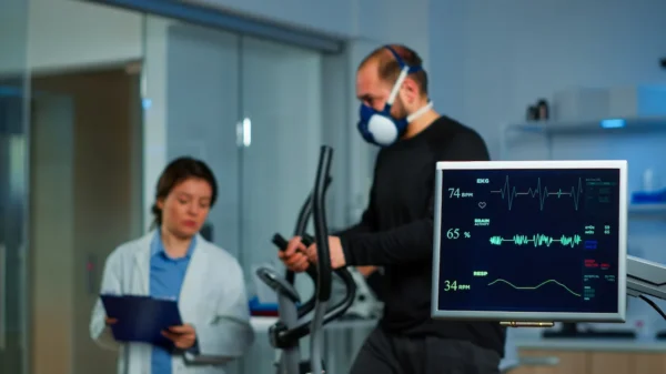
- Stress echocardiogram combines echocardiography with exercise. Images are taken at rest, then you exercise on a treadmill or stationary bike, and images are taken immediately after. This shows how your heart responds to increased demand and can reveal blockages in coronary arteries.
- Three-dimensional echocardiogram creates 3D images of heart structures rather than the standard 2D images. This provides better views of valves and helps surgeons plan procedures.
Why You Might Need This
Many situations lead doctors to order echocardiograms.
- Shortness of breath, particularly during activity or when lying flat, suggests possible heart failure or valve problems. The echocardiogram shows whether your heart is pumping adequately and whether valves are working properly.
- Chest pain can come from many causes. Echocardiograms help determine whether heart problems are responsible by showing how well your heart pumps and whether wall motion is normal.
- Heart murmurs heard through a stethoscope indicate turbulent blood flow, often from valve problems. Echocardiograms show exactly what’s wrong with the valve—whether it’s leaking, narrowed, or both—and how severe the problem is.
- After heart attacks, echocardiograms assess damage. They show which walls aren’t moving properly, how much the heart attack affected overall pumping, and whether complications like blood clots or valve damage occurred.
- Known heart conditions require periodic monitoring. If you have heart failure, valve disease, or cardiomyopathy, regular echocardiograms track whether your condition is stable, improving, or worsening.
- Before certain surgeries, particularly in people with risk factors, echocardiograms assess heart function to determine surgical risk.
- Abnormal electrocardiograms or chest X-rays sometimes prompt echocardiograms to see what’s causing the abnormalities.
- Family history of heart disease, particularly cardiomyopathy or congenital defects, might lead to screening echocardiograms.
Preparing for the Test
Standard transthoracic echocardiograms require no special preparation. Eat and drink normally. Take all your regular medications. Wear comfortable clothing that’s easy to remove from the waist up.
For transesophageal echocardiograms, don’t eat or drink anything for at least 6 hours before the test. You need an empty stomach because the tube going down your throat can trigger gagging or vomiting. Arrange for someone to drive you home since you’ll receive sedation.
For stress echocardiograms, wear comfortable clothes and athletic shoes suitable for exercise. Your doctor might ask you to stop certain heart medications before the test so they can see how your heart responds to stress without medication effects.
Bring a list of your medications and any previous test results. This helps doctors interpret your current study by comparing it to past ones.
What Happens During the Test
For a standard transthoracic echocardiogram, you’re taken to a dimly lit room—the darkness helps the technician see the screen better. You lie on an exam table, typically on your left side.
- Small electrocardiogram electrodes are placed on your chest to monitor your heart rhythm during the test.
- The technician applies gel to your chest. This gel helps sound waves travel from the transducer through your skin to your heart. It feels cool and slightly slippery.
- The transducer—a handheld device about the size of a computer mouse—is placed on your chest and moved around to different positions. The technician presses fairly firmly, which can be uncomfortable, particularly if you’re thin or have sensitive ribs. They’re trying to find “windows”—positions where they can see your heart clearly between ribs.
- You’ll be asked to hold your breath for a few seconds during some measurements. This helps get clearer images by keeping your lungs still.
- The technician is mostly silent during the test, concentrating on capturing good images. Don’t interpret silence as bad news—they’re focused on their work. A doctor will review the images and discuss results with you.
- The entire test takes 30-45 minutes. Afterward, the gel is wiped off and you can dress and leave immediately.
For transesophageal echocardiograms, an IV is placed and sedation is given. Your throat is numbed with spray. A tube with the transducer on its tip is gently passed down your throat into your esophagus. You’re sedated enough that you won’t remember much about this. The tube stays in place for 10-20 minutes while images are captured, then is removed. You’ll stay for observation until sedation wears off.
For stress echocardiograms, images are captured at rest first. Then you exercise on a treadmill or bike until you’re breathing hard and your heart is beating fast. Immediately after stopping—within seconds—you lie back down and more images are captured while your heart is still working hard.
Understanding Your Results
Echocardiogram reports describe multiple aspects of your heart’s structure and function.
- Chamber sizes indicate whether any chambers are enlarged. Enlarged left ventricles suggest high blood pressure or weak heart muscle. Enlarged left atria suggest long-standing valve problems or atrial fibrillation. Enlarged right chambers suggest lung problems or certain valve issues.
- Ejection fraction is the percentage of blood pumped out of the left ventricle with each beat. Normal is 55-70%. Below 40% indicates significant heart weakness. Between 40-50% is mildly reduced. Above 70% can indicate thickened, stiff heart muscle.
- Wall motion describes how each segment of heart muscle contracts. Normal means all segments thicken and move inward with each beat. Hypokinetic means a segment moves weakly. Akinetic means it doesn’t move. Dyskinetic means it bulges outward instead of contracting—this indicates dead muscle from a heart attack.
- Wall thickness shows whether muscle is normal, thickened, or thinned. Thickening occurs with high blood pressure or certain cardiomyopathies. Thinning occurs in areas damaged by heart attacks.
- Valve function is described for each of the four valves. Regurgitation means the valve leaks—blood flows backward when it should be sealed. Stenosis means the valve is narrowed—the opening is smaller than normal. Both can be graded as mild, moderate, or severe.
- Diastolic function describes how well your heart relaxes and fills between beats. Diastolic dysfunction means the heart is stiff and doesn’t fill properly, even though it might squeeze normally.
- Other findings might include fluid around the heart, blood clots inside chambers, tumors, or congenital defects.
- Your doctor explains what the findings mean for you specifically and what treatment, if any, is needed.
Limitations
Echocardiograms are excellent tests but have limitations.
- Image quality varies based on body habitus. Obesity, emphysema, or chest wall deformities can make it difficult to get clear images through the chest wall. Sometimes transesophageal studies are needed when transthoracic images are inadequate.
- Coronary arteries themselves aren’t well visualized. Echocardiograms show the effects of coronary disease—wall motion abnormalities and weak pumping—but don’t directly show blockages in the arteries. Coronary angiography or CT scans are needed to visualize the arteries themselves.
- Small abnormalities can be missed. Tiny valve vegetations from infections or small blood clots might not be visible, particularly on transthoracic studies.
- Measurements have some variability. Two different technicians or two different studies on the same day might get slightly different measurements. This is why trends over multiple studies are more meaningful than single measurements.
- Despite these limitations, echocardiography remains one of the most valuable cardiac tests because it’s safe, non-invasive, widely available, and provides comprehensive information about heart structure and function.
Key Points
- Echocardiograms use ultrasound to create moving images of your heart, showing chambers, valves, walls, and blood flow in real-time.
- The test is completely painless and safe with no radiation exposure. A transducer is placed on your chest and moved around to capture images from different angles.
- Standard transthoracic echocardiograms require no preparation. You can eat normally and take all medications. The test takes 30-45 minutes.
- Echocardiograms diagnose heart failure by showing weak pumping, valve problems by visualizing leaking or narrowed valves, heart attack damage by showing wall segments that don’t move properly, and many other conditions.
- Key measurements include ejection fraction showing how much blood your heart pumps with each beat, chamber sizes, wall thickness, valve function, and blood flow patterns.
- Normal ejection fraction is 55-70%. Below 40% indicates significant heart weakness. Chamber enlargement, valve problems, or wall motion abnormalities indicate specific heart conditions.
- Transesophageal echocardiograms provide more detailed images by placing the transducer down your throat. These require sedation and fasting beforehand.
- Stress echocardiograms combine imaging with exercise to show how your heart responds to increased demand.
- Results guide treatment decisions—whether you need medications, procedures to fix valves, or other interventions. Regular echocardiograms monitor known conditions over time.
- The test has limitations including variable image quality in some patients and inability to directly visualize coronary arteries themselves, but it remains one of the most valuable and widely used cardiac tests.
Reference: Echocardiogram